OrganRegen-SB202190 (FHPI)(C152121)

SB202190 (FHPI)

货号:C152121

规格:50mg

品牌:OrganRegen

产品介绍

DESCRIPTION

Background


SB202190 is a selective p38 MAP kinase inhibitor with IC50s of 50 nM and 100 nM for p38α and p38β2, respectively. SB 202190 binds to the ATP pocket of the active recombinant human p38 kinase with a Kd of 38 nM. SB 202190 has anti-cancer activity and rescued memory deficits[1][2]. SB202190 induces autophagy[3]. 

M. W t

331.34

Formula

C20H14FN3O

CAS No

152121-30-7

Storage

Powder

– 20°C

3 years

4°C

2 year                                  C20H14FN3O

In solvent

-80°C

6 months                                     

-20°C 

1 month

Solubility

DMSO

100 mg/mL(301.80 mM; Need ultrasonic)

Ethanol

12 mg/mL(36.22 mM)

H2O

< 0.1 mg/mL(insoluble)

OrganRegen-SB202190 (FHPI)(C152121)


技术参数

BIOLOGICAL ALTIVITY

In Vitro

SB202190 (0-10 μM; 0-72 hours) attenuates growth of a subgroup of CRC cell lines such as RKO, CACO2 and SW480 in a dose- and time-dependent manner[1].

SB 202190 strongly inhibited colony formation and anchorage-independent growth (10 μM for 7–10 days) and elevated apoptotic cell death (10 μM for 72 h) in this same subset of CRC lines (RKO, CACO2 and SW480)[2].

In RKO, CACO2 and SW480 cells, SB202190 (10 μM; 2 hours) abrogates phosphorylation of S6K1(T389) and S6(S235/236), but not AKT(S473), indicating that p38i selectively blocks mTORC1 signaling[2].

In Vivo

SB202190 (5 mg/kg; intraperitoneal injection; daily for 10-12 days) shows inhibition of tumor cell survival and tumor growth[2] .

REFERENCES
[1]. Davies SP, et al. Specificity and mechanism of action of some commonly used protein kinase inhibitors. Biochem J. 2000 Oct 1;351(Pt 1):95-105.

[2]. Nemoto S, et al. Induction of apoptosis by SB202190 through inhibition of p38beta mitogen-activated protein kinase. J Biol Chem. 1998 Jun 26;273(26):16415-20.

[3]. Grossi V, et al. Bay 43-9006 inhibits p38α activity in colorectal cancer cells and synergizes with the DFG-in inhibitor SB202190 to increase apoptotic response. Cancer Biol Ther. 2012 Dec;13(14):1471-81.

OrganRegen-Forskolin(C665752)

Forskolin

货号:C665752

规格:50mg

品牌:OrganRegen

产品介绍

OrganRegen-Forskolin(C665752)

DESCRIPTION

Background

Forskolin (Coleonol) is a potent adenylate cyclase activator with an IC50 of 41 nM and an EC50 of 0.5 μM for type I adenylyl cyclase[1]. Forskolin is also an inducer of intracellular cAMP formation[2]. Forskolin induces differentiation of various cell types and activates pregnane X receptor (PXR) and FXR[3]. Forskolin exerts a inotropic effect on the heart, and has platelet antiaggregatory and antihypertensive actions. Forskolin also induces autophagy[1][2][3].

Alias

毛喉素; Coleonol; Colforsin

M. W t

410.50

Formula

C22H34O7

CAS No

66575-29-9

Storage

Powder

-20°C  

3 years                          

In solvent

-80°C 

6 months                                 

-20°C      

1 month

Solubility

DMSO

100 mg/mL(243.61 mM; Need ultrasonic)

Ethanol

82 mg/mL(199.76 mM)

H2O

< 0.1 mg/mL(insoluble)


技术参数

BIOLOGICAL ALTIVITY

In Vitro  

Forskolin (Fsk) is a naturally occurring diterpene isolated from Coleus forskholii, directly activates adenylyl cyclase (AC) through its catalytic subunit to increase intracellular levels of cyclic adenosine monophosphate (cAMP)[1].

Forskolin (Fsk) affects the proliferation of the human T-cell lines such as Kit 225 and MT-2. Forskolin treatment inhibits the proliferation of both Kit 225 and MT-2 cells in a dose-dependent manner with an IC50 equal to ~5 μM Fsk. Forskolin treatment (10-100 μM) increases cAMPi levels ~5- to 20-fold above basal levels, which reache maximum levels between 50-100 μM Forskolin[4].

In Vivo  

The Forskolin (Coleonol)-treated Mrp4-/- mice shows an increased number of Ki67-positive and cleaved caspase 3-positive ECs, a significant decrease in the amount of pericyte coverage, and a reduced number of empty sleeves. In pups exposed to hyperoxia (75% oxygen) from P7 to P12, the Mrp4-/- mice shows a significant increase in the unvascularized retinal area[2].

REFERENCES

[1]. Robbins JD, et al. Forskolin carbamates: binding and activation studies with type I adenylyl cyclase. J Med Chem. 1996 Jul 5;39(14):2745-52.

[2]. Matsumiya W, et al. Forskolin modifies retinal vascular development in Mrp4-knockout mice. Invest Ophthalmol Vis Sci. 2012 Dec 7;53(13):8029-35.

[3]. Mayati A, et al. Functional polarization of human hepatoma HepaRG cells in response to forskolin. Sci Rep. 2018 Oct 31;8(1):16115.

[4]. Ríos-Silva M, et al. Effect of chronic administration of forskolin on glycemia and oxidative stress in rats with and without experimental diabetes. Int J Med Sci. 2014 Mar 11;11(5):448-52.

OrganRegen-DAPT(C208255)

DAPT

货号:C208255

规格:20mg

品牌:OrganRegen

产品介绍

DESCRIPTION

Background

DAPT (GSI-IX) is a potent and orally active γ-secretase inhibitor with IC50s of 115 nM and 200 nM for total amyloid-β (Aβ) and Aβ42, respectively. DAPT inhibits the activation of Notch 1 signaling and induces cell differentiation. DAPT also induces autophagy and apoptosis. DAPT has neuroprotection activity and has the potential for autoimmune and lymphoproliferative diseases, degenerative disease and cancers treatment[1][2].


OrganRegen-DAPT(C208255)


技术参数

Alias

GSI-IX

M. W t

432.46

Formula

C23H26F2N2O4

CAS No

208255-80-5

Storage

Powder

– 20°C

2 years

– 80°C

3 years

In solvent

– 80°C

12 months

– 20°C

3 months

Solubility

DMSO

≥ 100 mg/mL(283.71 mM)

Ethanol

41 mg/mL(94.81 mM)

H2O

< 0.1 mg/mL(insoluble)


BIOLOGICAL ALTIVITY

In Vitro  

DAPT inhibits Aβ production over 90%, effects only a modest reduction in APPβ in the culture media. Although APPβ is reduced by about 30% by DAPT treatment, this effect is not concentration-dependent and is reversed by the removal of DAPT[1]. CNE-2 cells are treated with increasing concentrations of DAPT (0, 25, 50 and 75 μM), and the γ-secretase-generated Notch 1 fragment Val1744-NICD is decreased after 48 h in a dose-dependent manner (P<0.01). The activation of γ-secretase is almost completely inhibited by DAPT at the concentration of 50 μM[3].

In Vivo 

DAPT is administered to PDAPP mice (100 mg/kg s.c.) and the levels of DAPT and Aβ are examined in the brain cortex. Peak DAPT levels of 490 ng/g are achieved in the brain 3 h after treatment, and levels greater than 100 ng/g (~200 nM) are sustained throughout the first 18 h. These brain concentrations of DAPT are in excess of its IC50 for lowering Aβ in neuronal cultures (115 nM), and results in a robust and sustains pharmacodynamic effect[1]. DAPT protects brain against cerebral ischemia by down-regulating the expression of Notch 1 and Nuclear factor kappa B in rats. Western blot analyses also show a significant decrease of Notch 1 and NF-κB expression in DAPT (0.03 mg/kg) group (P<0.05 vs. MCAO group)[2].


REFERENCES

[1]. Dovey HF, et al. Functional gamma-secretase inhibitors reduce beta-amyloid peptide levels in brain. J Neurochem. 2001 Jan;76(1):173-81.

[2]. Li S, et al. DAPT protects brain against cerebral ischemia by down-regulating the expression of Notch 1 and nuclear factor κB in rats. Neurol Sci. 2012 Dec;33(6):1257-64.

[3]. Zhou JX, et al. γ-secretase inhibition combined with NSC 119875 enhances apoptosis of nasopharyngeal carcinoma cells.Exp Ther Med. 2012 Feb;3(2):357-361.

OrganRegen-Dexamethasone (DHAP)(C500220)

Dexamethasone (DHAP)

货号:C500220

规格:10mM (in 1mL DMSO)

品牌:OrganRegen

产品介绍

DESCRIPTION

Background

Dexamethasone is a glucocorticoid receptor agonist. Dexamethasone also significantly decreases CD11b, CD18, and CD62L expression on neutrophils, and CD11b and CD18 expression on monocytes. Dexamethasone is highly effective in the control of COVID-19 infection. Dexamethasone inhibits production of exosomes containing inflammatory microRNA-155 in lipopolysaccharide-induced macrophage inflammatory responses.

Alias

Synonyms,地塞米松,Hexadecadrol,Prednisolone F

M. W t

392.46

Formula

C22H29FO5

CAS No

50-02-2

Storage

Powder

-20°C

3 years                                                                                                                             

In solvent

-80°C     

2 years                  

Solubility

DMSO 

250 mg/mL(637.01 mM; ultrasonic and warming and heat to 60°C)

Ethanol 

8.33 mg/mL(21.23 mM; Need ultrasonic)

H2O

< 0.1 mg/mL(insoluble)


技术参数

BIOLOGICAL ALTIVITY

In Vitro   

Dexamethasone regulates several transcription factors, including activator protein-1, nuclear factor-AT, and nuclear factor-kB, leading to the activation and repression of key genes involved in the inflammatory response[1].

Dexamethasone potently inhibits granulocyte-macrophage colony stimulating factor (GM-CSF) release from A549 cells with EC50 of 2.2 nM. Dexamethasone (EC50=36 nM) induces transcription of the β2-receptor is found to correlate with glucocorticoid receptor (GR) DNA binding and occurred at 10-100 fold higher concentrations than the inhibition of GM-CSF release. Dexamethasone (IC50=0.5 nM) inhibits a 3×κB (NF-κB, IκBα, and I-κBβ), which is associated with inhibition of GM-CSF release[2].

In Vivo  

It has previously been reported that treatment with Dexamethasone at a dose of 2×5 mg/kg efficiently inhibits lipopolysaccharide (LPS)-induced inflammation. In our experimental system, treatment with a single dose of Dexamethasone 10 mg/kg (i.p.) significantly decreases recruitment of granulocytes as well as spontaneous production of oxygen radicals compared with animals expose to LPS and injected with solvent alone (saline). The effects are statistically significant when administered both 1 h before and 1 h after inhalation of LPS. The number of granulocytes in BALF decreased to levels comparable to healthy animals (given an aerosol of water)[3].

REFERENCES

[1]. LaLone CA, et al. Effects of a glucocorticoid receptor agonist, Dexamethasone, on fathead minnow reproduction, growth, and development. Environ Toxicol Chem. 2012 Mar;31(3):611-22.

[2]. Adcock IM, et al. Ligand-induced differentiation of glucocorticoid receptor (GR) trans-repression and transactivation: preferential targetting of NF-kappaB and lack of I-kappaB involvement. Br J Pharmacol. 1999 Jun;127(4):1003-11

[3]. Rocksén D, et al. Differential anti-inflammatory and anti-oxidative effects of Dexamethasone and N-acetylcysteine in endotoxin-induced lung inflammation. Clin Exp Immunol. 2000 Nov;122(2):249-56

OrganRegen-Hydrocortisone(C502370)

Hydrocortisone

货号:C502370

规格:500mg

品牌:OrganRegen

产品介绍

OrganRegen-Hydrocortisone(C502370)

DESCRIPTION

Background


Hydrocortisone is a steroid hormone or glucocorticoid secreted by the adrenal cortex[1].

Alias

Cortisol;氢化可的松;17-羟基皮质 (酮;皮质甾醇;氢化皮质素

M. W t

362.46

Formula

C21H30O5

CAS No

50-23-7

Storage

Powder

-20 °C          

3 years

In solvent

-80 °C   

6 months

-20 °C          

1 month                                       

Solubility

DMSO     

H2O : < 0.1 mg/mL

≥ 31 mg/mL(85.53 mM)


Ethanol

23 mg/mL(63.46 mM)

H2O

< 0.1 mg/mL(insoluble)

BIOLOGICAL ALTIVITY

In Vitro   

Hydrocortisone (50 nM) shows a dose-dependent down-regulation of GR transcript in hCMEC/D3 cells. Hydrocortisone supplementation of the serum-reduced cell differentiation medium leads to a significant increase in TER across the hCMEC/D3 monolayer[1]. Hydrocortisone-treated Dendritic cells (DCs) show a decreased expression of MHC II molecules, the costimulatory molecule CD86, and the DC-specific marker CD83, as well as a strongly reduced IL-12 secretion. Hydrocortisone-treated DCs inhibit production of IFN-γ but induce an increased release of IL-4 and no change in IL-5[2]. Hydrocortisone reduces postischemic oxidative stress, perfusion pressure, and transudate formation. Hydrocortisone inhibits postischemic shedding of syndecan-1, heparan sulfate, and hyaluronan as is release of histamine from resident mast cells[3].

In Vivo

NCT00621985

Boston Children´s Hospital

Adrenal Hyperplasia, Congenital

Phase 2

NCT03910088

Cairo University

Post-Dural Puncture Headache

Phase 4

NCT00657306

University of Turin, Italy

Cirrhosis With Ascites

Phase 2


REFERENCES

[1].Förster C, et al. Differential effects of hydrocortisone and TNFalpha on tight junction proteins in an in vitro model of the human blood-brain barrier. J Physiol. 2008 Apr 1;586(7):1937-49.

[2]. Bellinghausen I, et al. Inhibition of human allergic T-cell responses by IL-10-treated dendritic cells: differences from hydrocortisone-treated dendritic cells. J Allergy Clin Immunol. 2001 Aug;108(2):242-9.

[3].Chappell D, et al. Hydrocortisone preserves the vascular barrier by protecting the endothelial glycocalyx. Anesthesiology. 2007 Nov;107(5):776-84.

OrganRegen-CHIR-99021(C252917)

CHIR-99021

货号:C252917

规格:10mg

品牌:OrganRegen

产品介绍

DESCRIPTION

Background

CHIR-99021 is a potent and selective GSK-3α/β inhibitor with IC50s of 10 nM and 6.7 nM. Laduviglusib shows >500-fold selectivity for GSK-3 over CDC2, ERK2 and other protein kinases. Laduviglusib is also a potent Wnt/β-catenin signaling pathway activator. Laduviglusib enhances mouse and human embryonic stem cells self-renewal. Laduviglusib induces autophagy[1][2][3].

Alias

Laduviglusib; CT99021


M. W t

465.34

Formula

C22H18Cl2N8

CAS No

252917-06-9

Storage

Powder

-20°C

3 years

4°C

2 years                               C22H18Cl2N8

In solvent

-80°C

6 months         

-20°C

1 month

Solubility

DMSO       

16.67 mg/mL(35.82 mM)

H2O

< 0.1 mg/mL(insoluble)

BIOLOGICAL ALTIVITY

In Vitro  

CHIR-99021 inhibits human GSK-3β with Ki values of 9.8 nM[1]. Laduviglusib is a small organic molecule that inhibits GSK3α and GSK3β by competing for their ATP-binding sites.In vitro kinase assays reveal that Laduviglusib specifically inhibits GSK3β (IC50=~5 nM) and GSK3α (IC50=~10 nM), with little effect on other kinases[4]. In the presence of Laduviglusib the viability of the ES-D3 cells is reduced by 24.7% at 2.5 μM, 56.3% at 5 μM, 61.9% at 7.5 μM and 69.2% at 10 μM Laduviglusib with an IC50 of 4.9 mm[2].

In Vivo

In ZDF rats, a single oral dose of Laduviglusib (16 mg/kg or 48 mg/kg) rapidly lowers plasma glucose, with a maximal reduction of nearly 150 mg/dl 3-4 h after administration[1]. Laduviglusib (2 mg/kg) given once, 4 h before irradiation, significantly improves survival after 14.5 Gy abdominal irradiation (ABI). Laduviglusib treatment significantly blocks crypt apoptosis and accumulation of p-H2AX+ cells, and improves crypt regeneration and villus height. Laduviglusib treatment increases Lgr5+ cell survival by blocking apoptosis, and effectively prevents the reduction of Olfm4, Lgr5 and CD44 as early as 4 h[5].

REFERENCES

[1]. Ring DB, et al. Selective glycogen synthase kinase 3 inhibitors potentiate activation of glucose transport and utilization in vitro and in vivo. Diabetes. 2003 Mar;52(3):588-95.

[2]. Naujok O, et al. Cytotoxicity and activation of the Wnt/beta-catenin pathway in mouse embryonic stem cells treated with four GSK3 inhibitors.BMC Res Notes. 2014 Apr 29;7:273.

[3]. Ye S, et al. Pleiotropy of glycogen synthase kinase-3 inhibition by CHIR99021 promotes self-renewal of embryonic stem cells from refractory mouse strains. PLoS One. 2012;7(4):e35892.

[4]. Bennett CN, et al. Regulation of Wnt signaling during adipogenesis. J Biol Chem. 2002 Aug 23;277(34):30998-1004.

[5]. Wang X, et al. Pharmacologically blocking p53-dependent apoptosis protects intestinal stem cells and mice from radiation. Sci Rep. 2015 Apr 10;5:8566.

OrganRegen-Estradiol(C502820)

Estradiol

货号:C502820

规格:1g

品牌:OrganRegen

产品介绍

DESCRIPTION

Background

Estradiol is a steroid sex hormone vital to the maintenance of fertility and secondary sexual characteristics in females. Estradiol upregulates IL-6 expression through the estrogen receptor β (ERβ) pathway[1][2][3].


技术参数

Alias

β-Estradiol; E2; 17β-Estradiol; 17β-Oestradiol雌二醇;强力求偶素

M. W t

272.38

Formula

C18H24O2

CAS No

50-28-2

Storage

Powder

-20°C

3 years

4°C

2 years

In solvent

-80°C

6 months                                   

-20°C

1 month

Solubility

DMSO      

≥ 100 mg/mL(283.71 mM)

Ethanol

14 mg/mL(51.40 mM)

H2O

< 0.1 mg/mL(insoluble)

BIOLOGICAL ALTIVITY

In Vitro   

Estradiol causes new dendritic spines and synapses in hippocampal CA1 pyramidal cells. Estradiol increases NMDA receptor binding by 46% in parallel with dendritic spine and synapse density. Estradiol also elevates sensitivity of CA1 pyramidal cells to NMDA receptor-mediated synaptic input and such an effect is correlated with the estradiol-induced increase in dendritic spine density in the apical dendritic tree of these cells[1]. Estradiol reduces Ba2+ entry reversibly via Ca2+ channels in acutely dissociated and cultured neostriatal neurons. Estradiol also reduces Ba2+ currents but is significantly less effective than Estradiol in rat neostriatal neurons[2].

In Vivo  

Estradiol functions in hippocampal synapse density during the estrous cycle in the adult rat[4].Estradiol reverses the ovariectomy-induced decrease in spine density. Estradiol in combination with progesterone enhances spine density for 2 to 6 h but decreases following estradiol alone[5].Estradiol (0.2 mg/kg; i.p.; C57BL/6 female mice) plus 10 or 20 mg/kg progesterone, enhances memory consolidation in young ovariectomized mice[6].

REFERENCES

[1]. Förster C, et al. Differential effects of hydrocortisone and TNFalpha on tight junction proteins in an in vitro model of the human blood-brain barrier. J Physiol. 2008 Apr 1;586(7):1937-49.

[2]. Bellinghausen I, et al. Inhibition of human allergic T-cell responses by IL-10-treated dendritic cells: differences from hydrocortisone-treated dendritic cells. J Allergy Clin Immunol. 2001 Aug;108(2):242-9.

[3]. Chappell D, et al. Hydrocortisone preserves the vascular barrier by protecting the endothelial glycocalyx. Anesthesiology. 2007 Nov;107(5):776-84.

[4]. Woolley CS, et al. Estradiol mediates fluctuation in hippocampal synapse density during the estrous cycle in the adult rat. J Neurosci. 1992 Jul;12(7):2549-54.

[5]. Woolley CS, et al. Roles of estradiol and progesterone in regulation of hippocampal dendritic spine density during the estrous cycle in the rat. J Comp Neurol. 1993 Oct 8;336(2):293-306.

[6]. Harburger LL, et al. Dose-dependent effects of post-training estradiol plus progesterone treatment on object memory consolidation and hippocampal extracellular signal-regulated kinase activation in young ovariectomized mice. Neuroscience. 2009;160(1):6-12.

OrganRegen-Prostaglandin E2(C363246)

Prostaglandin E2

货号:C363246

规格:20mg

品牌:OrganRegen

产品介绍

DESCRIPTION

Background

Prostaglandin E2 is a hormone-like substance that participate in a wide range of body functions such as the contraction and relaxation of smooth muscle, the dilation and constriction of blood vessels, control of blood pressure, and modulation of inflammation.

Alias

PGE2;前列腺素 E2;地诺前列酮


技术参数

M. W t

352.47

Formula

C₂₀H₃₂O₅

CAS No

363-24-6

Storage

Powder

-20°C  3 years         

In solvent

-80°C  6 months

-20°C  1 month                             

Solubility

DMSO    

≥ 100 mg/mL(283.71 mM)

Ethanol

70 mg/mL(198.60 mM)

H2O

< 0.1 mg/mL(insoluble)

BIOLOGICAL ALTIVITY

In Vitro   

PGE2 shows inhibition of IL 2 production in the mixture of irradiated and nonirradiated T lymphocytes. PGE2 (0.1-10 μM) dose-dependently inhibits the production of IL 2. PGE2 acts during the inductive phase of activation of suppressor cells. Preincubation of T lymphocytes with PGE2 induces cells that suppress IL 2 production and PHA proliferation[1].

In Vivo  

PGE2 (0.3 μg/k, i.p.) significantly reduces the number of peritoneab macrophages undergoing phagocytosis of the methacrybate microbeads in rats[2]. PGE2 (0.1 mg/min, i.a.) increases renal blood flow. PGE2 produces a biphasic change in renal vascular resistance, vasodilatation starts at 0.01 mg/min and is maximal at about 3 mg/min, while at the highest dose used (20 mg/min) PGE2 induces renal vasoconstriction[3].

REFERENCES

[1]. Chouaib S, et al. The mechanisms of inhibition of human IL 2 production. II. PGE2 induction of suppressor T lymphocytes. J Immunol. 1984 Apr;132(4):1851-7.

[2]. Fernandez-Repollet E, et al. In vivo effects of prostaglandin E2 and arachidonic acid on phagocytosis of fluorescent methacrylate microbeads by rat peritoneal macrophages. J Histochem Cytochem. 1982 May;30(5):466-70.

[3]. Haylor J, et al. Renal vasodilator activity of prostaglandin E2 in the rat anaesthetized with pentobarbitone. Br J Pharmacol. 1982 May;76(1):131-7.

OrganRegen-Y-27632 dihydrochloride(C629401)

Y-27632 dihydrochloride

货号:C629401

规格:50mg

品牌:OrganRegen

产品介绍

DESCRIPTION

Background


Y-27632 dihydrochloride is an orally active, ATP-competitive inhibitor of ROCK-I and ROCK-II, with Kis of 220 and 300 nM, respectively. Y-27632 dihydrochloride attenuates Doxorubicin-induced apoptosis of human cardiac stem cells. Y-27632 also suppresses dissociation-induced apoptosis of murine prostate stem/progenitor cells. Y-27632 dihydrochloride primes human induced pluripotent stem cells (hIPSCs) to selectively differentiate towards mesendodermal lineage via epithelial-mesenchymal transition-like modulation[1][2][3][4][5].


技术参数

M. W t

320.26

Formula

C14H23Cl2N3O

CAS No

129830-38-2                                                

Storage

Powder

-20 °C

3 years

In solvent

-80°C

6 months

-20 °C

1 month

Solubility

DMSO    

33.33 mg/mL(104.07 mM; Need ultrasonic)

H2O

100 mg/mL(312.25 mM; Need ultrasonic)

BIOLOGICAL ALTIVITY

In Vitro   

Y-27632 inhibits the ROCK family of kinases 100 times more potently than other kinases including protein kinase C, cAMP-dependent kinase and myosin light chain kinase. Y-27632 prolongs the lag time and delays the appearance of BrdU-labeled cells in a concentration-dependent manner, delays of about 1 and 4 h are noticed in the Swiss 3T3 cells treated with 10 and 100 μM Y-27632, respectively[1].
Y-27632 promotes neuronal differentiation of adipose tissue-derived stem cells (ADSCs). Compared to 1.0 and 2.5 μM Y-27632 induced groups, percentages of neuroal-like cells achieved a peak in the 5.0 μM Y-27632 induced group[2].

In Vivo  

Y-27632 (5 and 10 mg/kg) significantly prolongs the onset time of myoclonic jerks when compare with saline group. Y-27632 (5 and 10 mg/kg) significantly prolongs the onset time of clonic convulsions when compare with saline group[3].
Treatment with Dimethylnitrosamine (DMN) causes a significant decrease in rat body and liver weight (DMN-S group) compared with control animals (S-S group). Oral Y27632 (30 mg/kg) essentially prevents this DMN-induced rat body and liver weight loss (DMN-Y group)[4].

REFERENCES

[1]. Ishizaki T, et al. Pharmacological properties of Y-27632, a specific inhibitor of rho-associated kinases. Mol Pharmacol. 2000 May;57(5):976-83.

[2]. Xue ZW, et al. Rho-associated coiled kinase inhibitor Y-27632 promotes neuronal-like differentiation of adult human adipose tissue-derived stem cells.Chin Med J (Engl). 2012 Sep;125(18):3332-5.

[3]. Tada S, et al. A selective ROCK inhibitor, Y27632, prevents dimethylnitrosamine-induced hepatic fibrosis in rats. J Hepatol. 2001 Apr;34(4):529-36.

[4]. Inan S, et al. Antiepileptic effects of two Rho-kinase inhibitors, Y-27632 and fasudil, in mice. Br J Pharmacol. 2008 Sep;155(1):44-51.

[5]. Maldonado M, et al. ROCK inhibitor primes human induced pluripotent stem cells to selectively differentiate towardsmesendodermal lineage via epithelial-mesenchymal transition-like modulation. Stem Cell Res. 2016 Sep;17(2):222-227.


OrganRegen-Recombinant Human R-spondin1(861-RS1)

Recombinant Human R-spondin1

货号:861-RS1

规格:1mg

100ug

品牌:OrganRegen

产品介绍

Background:

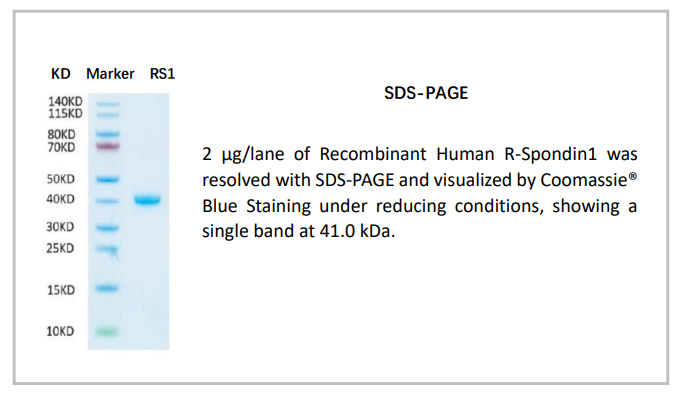
R-Spondin1 (Rspo-1) belongs to the (Rspo) family of Wnt modulators. Currently, the family consists of four structurally related secreted ligands (Rspo 1-4), all containing the furin-like and thrombospondin structural domains. Postnatally, R-Spondin1 is expressed by neuroendocrine cells in the intestine, adrenal gland and pancreas, and by epithelia in kidney and prostate. R-Spondin1 regulates Wnt/β-catenin by competing with the Wnt antagonist DKK-1 for binding to the Wnt coreceptors, Kremen and LRP-6, reducing their DKK-1-mediated internalization. Recombinant Human R-Spondin1 is a 27.3 kDa protein consisting of 233 amino acid residues. Due to glycosylation, RSpondin1 migrates at an apparent molecular weight of approximately 41.0 kDa by SDS-PAGE analysis under reducing conditions.


技术参数

Source:

Chinese Hamster Ovary cell line

Protein Construction:

A DNA sequence encoding the amino acids (Ala31-Ser263) of human Rspo-1 (Accession Number: Q2MKA7) was expressed.

Synonyms:

Roof plate-specific spondin-1, RSPO1

Purity:

≥ 95%, by SDS-PAGE visualized with quantitative densitometry by Coomassie® Blue Staining.

Biological Activity:

Measured by its ability to induce activation of β-catenin response in a Top-flash Luciferase assay using HEK293T human embryonic kidney cells. The ED50 for this effect is typically 0.1-0.5 μg/ml in the presence of 5 ng/ml recombinant mouse Wnt3a.

Endotoxin Level:

<0.10 EU per 1 μg of the protein by the LAL method

Calculated Molecular Weight:

27.3 kDa

SDS-PAGE:

41.0 kDa, reducing condition

OrganRegen-Recombinant Human R-spondin1(861-RS1)

Formulation:

The product is Lyophilized from a 0.22μm filtered solution in PBS.

Shipping:

The product is shipped on ice. Upon receipt, store it immediately as methods recommended below.

Reconstitution:

Reconstitute in sterile PBS buffer containing 0.1 % BSA to a concentration of 0.1-1.0 mg/mL.

Stability & Storage:

24 months, -20 to -70 °C, under powder state; 12 months, -20 to -70 °C, under sterile conditions after reconstitution; 2 month, 2 to 8 °C under sterile conditions after reconstitution; avoid repeated freeze-thaw cycles.

References:

  1. Kim, K.A., et al., R-Spondin proteins: a novel link to beta-catenin activation. Cell Cycle, 2006. 5(1)

  2. Kamata, T., et al., R-spondin, a novel gene with thrombospondin type 1 domain, was expressed in the dorsal neural tube and affected in Wnts mutants. Biochim Biophys Acta, 2004. 1676(1)

  3. Hao, H.X., et al., ZNRF3 promotes Wnt receptor turnover in an R-spondin-sensitive manner. Nature, 2012. 485(7397)